
Upwell’s Dr. Heather Harris gives us an inside look at the Advanced Veterinary Techniques workshop at the 2019 International Sea Turtle Symposium in Charleston, South Carolina. From her perspective as a veterinary specialist, Dr. Harris shares insights on cutting-edge applications and tools to assess the health of endangered sea turtles.

Have you ever wondered how to tell whether an immature sea turtle is male or female? Or if an adult female sea turtle is preparing to lay eggs? Or whether a sea turtle with visible external fibropapilloma tumors also has hidden tumors on her internal organs? Or if she may suffer from gas bubble disease as a result of fisheries bycatch? The tools of diagnostic imaging can help answer all of these questions. Veterinarians are trained to use imaging (radiography, ultrasound, CT, MRI, and endoscopy) in clinical practice to diagnose disease and monitor response to treatment. The comparative nature of veterinary medicine teaches us to apply these techniques in diverse species from mammals to reptiles to birds to fish. These same tools are used by wildlife vets to assess the health of endangered sea turtles and facilitate research to aid in population management and recovery.
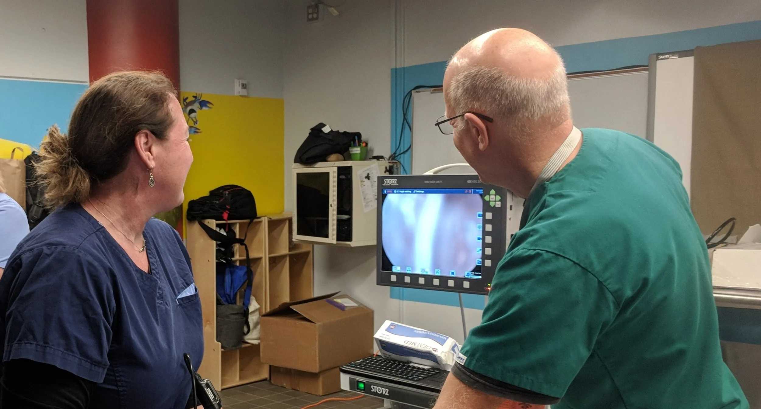
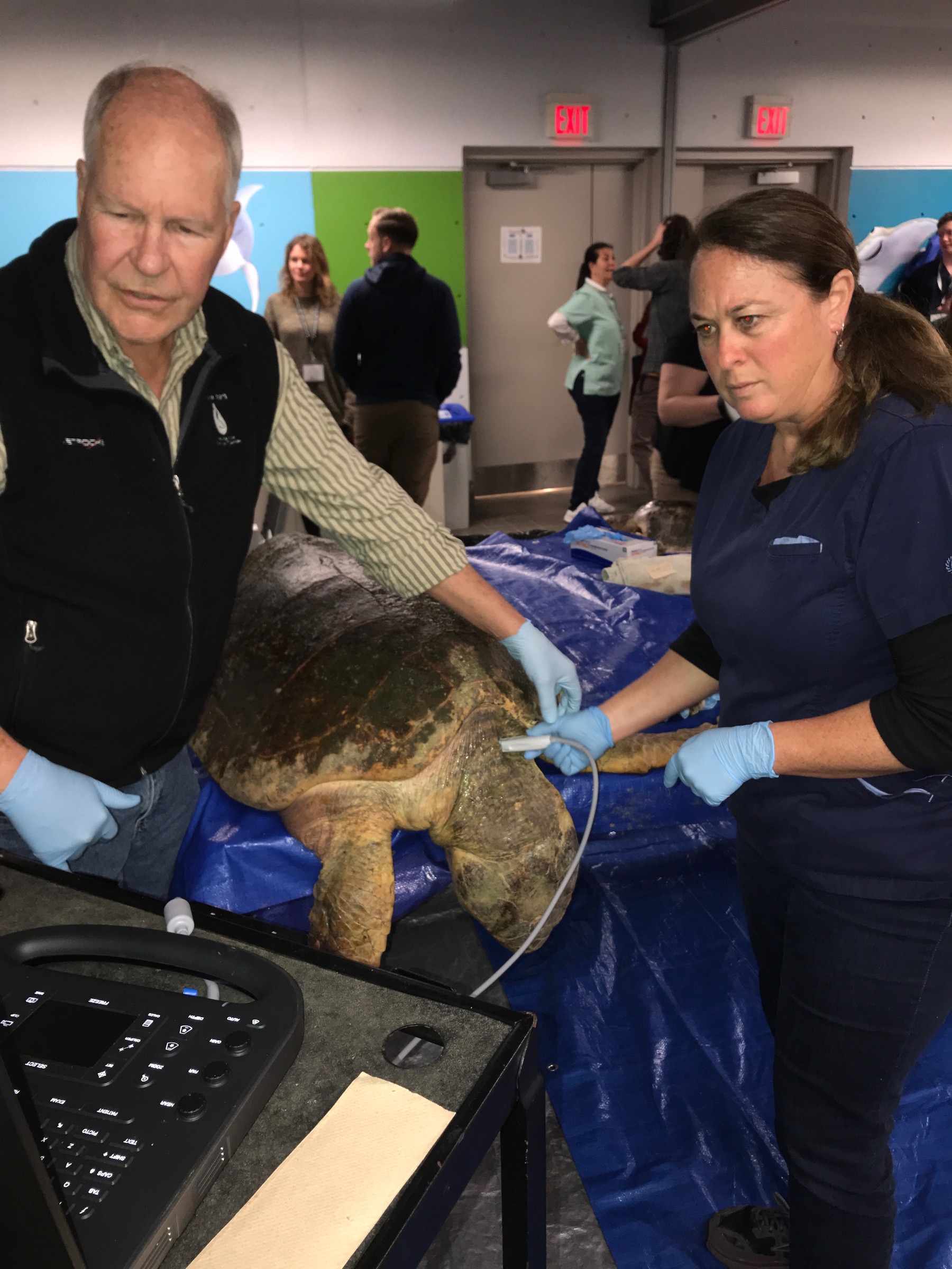
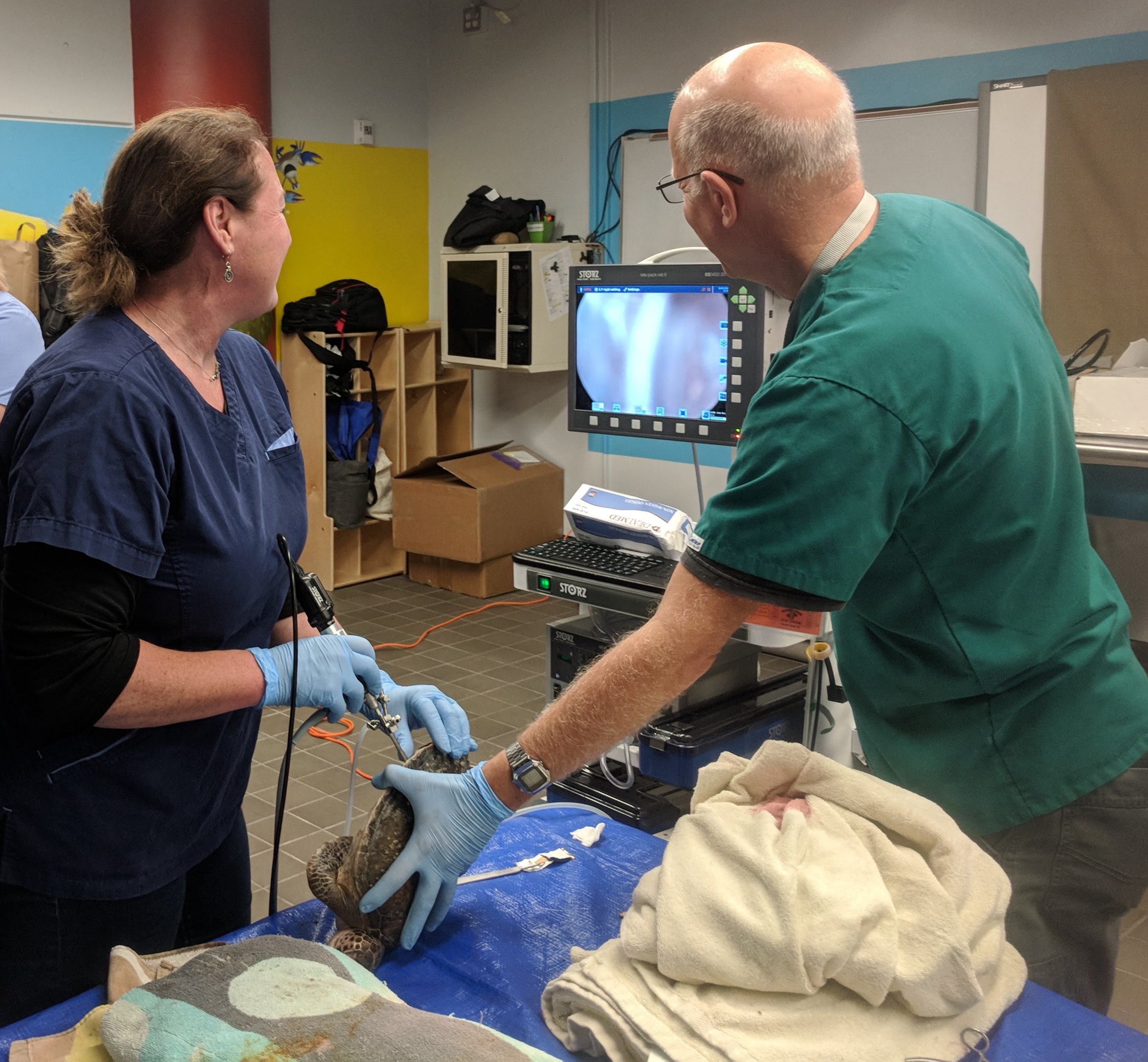
So, when you invite a group of highly enthusiastic (and slightly geeky) sea turtle veterinarians and technicians to visit a state-of-the-art aquarium clinic filled with fancy diagnostic imaging tools and multiple species of sea turtle carcasses of various sizes, a happy orderly chaos ensues. This is exactly what occurred this past weekend at the Advanced Veterinary Techniques Workshop as part of the International Sea Turtle Symposium in Charleston, SC. Vets and techs from all over the world who work on sea turtles converged at the South Carolina Aquarium to learn from experts in the field, experiment with new equipment, and exchange ideas and experiences. The room was a dynamic flow with clusters of people forming spontaneously around hands-on demonstrations and lively debates igniting on such topics as the optimal location to insert the endoscope, favorite obscure blood collection sites, preferences for anesthetic drug combinations, surgical approaches for fishing hook removal, and how to administer an epidural. With a CT machine on site, we were able to obtain images from patients and review cases where CT had provided the level of detail needed to confirm the diagnosis. We learned about cross-matching sea turtles for blood transfusions using simple techniques and equipment available in most clinics. And we also had the unique opportunity to observe the surgical removal of external tumors using laser technology in a green turtle with advanced stage fibropapillomatosis, a devastating disease preferentially affecting green turtles in warm water. It was a fantastic and highly informative event that will certainly improve our ability to provide the best achievable care for our patients in rehabilitation and field research around the world.
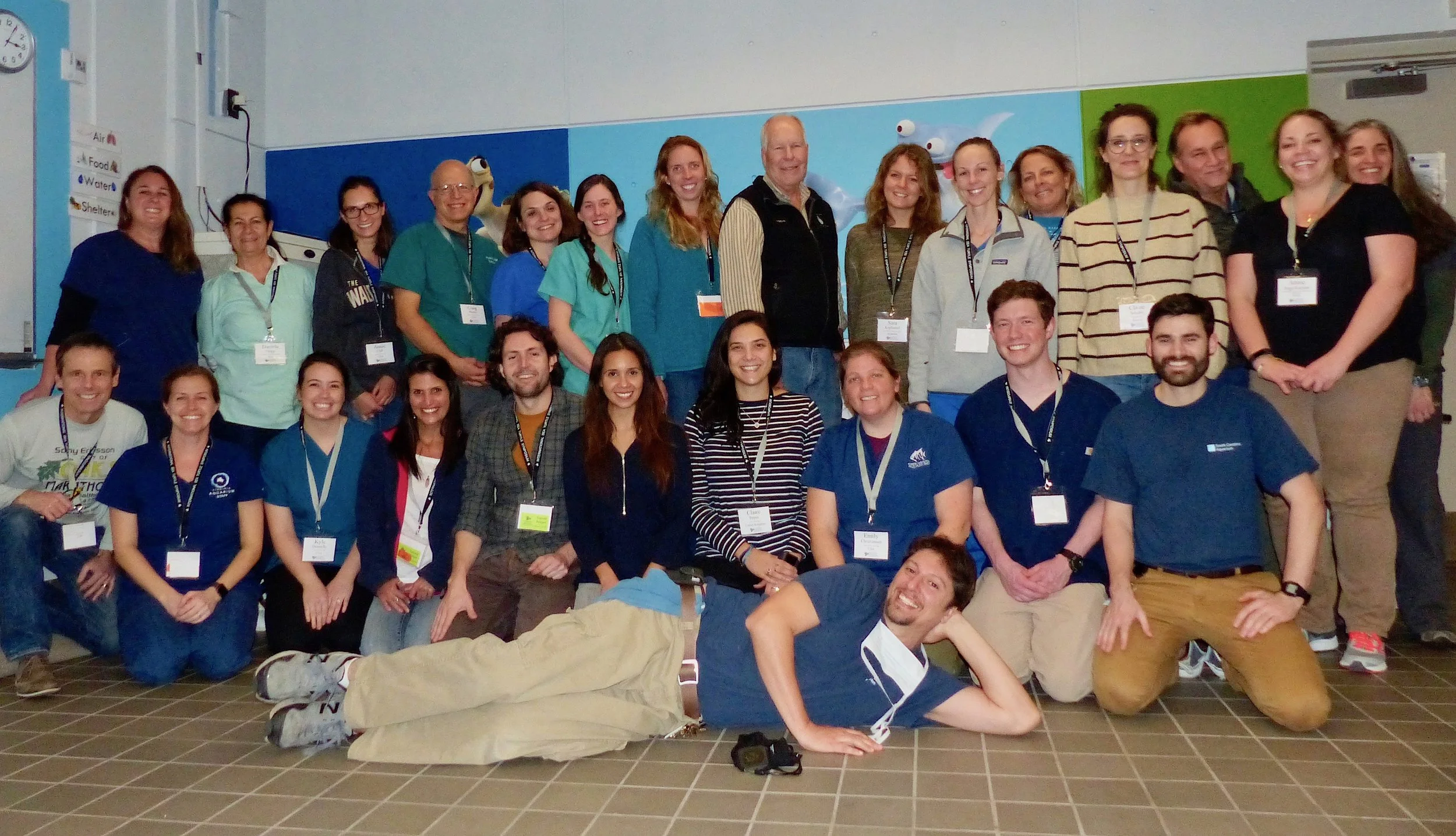
A huge thank you to Dr. Shane Boylan and the staff from the South Carolina Aquarium for organizing and hosting this excellent workshop and offering continuing education credit. Additional thanks to instructors Dr. Craig Harms and Dr. Greg Lewbart from the North Carolina State University, Dr. Terry Norton from the Georgia Sea Turtle Center, Dr. Bob George retired vet from the Virginia Aquarium, Dr. Emily Christiansen from the North Carolina Aquariums, Dr. Kyle Donnelly from the University of Florida, and sales reps from Sonosite and Storz for providing equipment and lunch. And an enormous thank you to Upwell for generously supporting my involvement in this workshop.
Heather S. Harris, DVM, MPVM, DACVPM
Wildlife Veterinarian